
ULM: Ultrasound Localization Microscopy
In the 1880s, the Curie brothers first described piezoelectric sound waves, laying the foundation for modern sonography (Curie et al. 1880). Although “ultrasound waves” initially served military purposes (echolocation), they successfully entered medical practice from the mid-20th century onwards (Dussik 1942, Donald et al. 1958). To this day, B-mode sonography is characterised in particular by the precise morphological imaging of tissues, although its interpretation largely depends on the investigator. Sonography holds a particularly significant place in paediatrics. Its non-invasive, rapid, and targeted diagnostics make it the method of choice for many clinical questions (Regensburger et al. 2019). Newer methods, such as Acoustic Radiation Force Impulse (ARFI) elastography, also permit quantitative analyses, for example measuring liver stiffness as a surrogate parameter for liver fibrosis in patients with cystic fibrosis (Schnell et al. 2023). The use of intravenously administered contrast agents allows further, sometimes highly complex issues to be resolved in paediatric sonography (Knieling et al. 2016). For instance, off-label use in infants has enabled transfontanelle imaging and quantification of cerebral perfusion during complex paediatric cardiac surgery on the heart-lung machine (Knieling et al. 2020, Knieling et al. 2022, Rüffer et al. 2022) (video). New possibilities in AI-assisted evaluation and tracking of individual microbubbles enable unique “super-resolution imaging” (Ultrasound-Localization Microscopy, ULM) (Errico et al. 2015). This can be used, for example, to characterise the smallest vessels in newborns with stroke or following vascular interventions (video).
Hoerning, A., J. Jüngert, G. Siebenlist, F. Knieling and A. P. Regensburger (2024). “Ultrasound in Pediatric Inflammatory Bowel Disease-A Review of the State of the Art and Future Perspectives.” Children (Basel) 11(2)
Danko, V., J. Jüngert, S. Schuessler, A. Buehler, D. Klett, A. Federle, A. Roos, H. Lochmüller, M. F. Neurath, J. Woelfle, R. Trollmann, M. J. Waldner, F. Knieling, A. P. Regensburger and A. L. Wagner (2023). “Hybrid reflected-ultrasound computed tomography versus B-mode-ultrasound for muscle scoring in spinal muscular atrophy.” J Neuroimaging 33(3)
Schnell, A., J. Jüngert, D. Klett, H. Hober, N. Kaiser, R. Ruppel, A. Geppert, C. Tremel, J. Sobel, E. Plattner, S. Schmitt-Grohé, S. Zirlik, D. Strobel, M. F. Neurath, F. Knieling, M. Rauh, J. Woelfle, A. Hoerning and A. P. Regensburger (2023). “Increase of liver stiffness and altered bile acid metabolism after triple CFTR modulator initiation in children and young adults with cystic fibrosis.” Liver Int 43(4)
Knieling, F., R. Cesnjevar, A. P. Regensburger, A. L. Wagner, A. Purbojo, S. Dittrich, F. Münch, A. Neubert, J. Woelfle, J. Jüngert and A. Rüffer (2022). “Transfontanellar Contrast-enhanced US for Intraoperative Imaging of Cerebral Perfusion during Neonatal Arterial Switch Operation.” Radiology 304(1)
Rüffer, A., F. Knieling, R. Cesnjevar, A. Regensburger, A. Purbojo, S. Dittrich, F. Münch, J. Wölfle and J. Jüngert (2022). “Equal cerebral perfusion during extended aortic coarctation repair.” Eur J Cardiothorac Surg 61(2)
Aguilar, C., A. P. Regensburger, F. Knieling, A. L. Wagner, G. Siebenlist, J. Woelfle, H. Koehler, A. Hoerning and J. Jüngert (2021). “Pediatric Buried Bumper Syndrome: Diagnostic Validity of Transabdominal Ultrasound and Artificial Intelligence.” Ultraschall Med 43(05)
Knieling, F., A. Rüffer, R. Cesnjevar, A. P. Regensburger, A. Purbojo, S. Dittrich, F. Münch, A. Neubert, S. Meyer, D. Strobel, W. Rascher, J. Woelfle and J. Jüngert (2020). “Transfontanellar Contrast-Enhanced Ultrasound for Monitoring Brain Perfusion During Neonatal Heart Surgery.” Circ Cardiovasc Imaging 13(3)
Regensburger, A. P., F. Knieling, A. Feldkamp, W. Rascher, K. Diesch, J. Woelfle, H. U. Prokosch and J. Jungert (2019). “Time Tracking of Standard Ultrasound Examinations in Pediatric Hospitals and Pediatric Medical Practices – A Multicenter Study by the Pediatric Section of the German Society of Ultrasound in Medicine (DEGUM).” Ultraschall Med
Regensburger, A. P., A. L. Wagner, G. Hanslik, S. C. Schussler, F. B. Fahlbusch, J. Woelfle, J. Jungert, R. Trollmann and F. Knieling (2019). “Ultra-high-frequency ultrasound in patients with spinal muscular atrophy: A retrospective feasibility study.” Muscle Nerve
Knieling, F., D. Strobel, O. Rompel, M. Zapke, C. Menendez-Castro, M. Wolfel, J. Schulz, W. Rascher and J. Jungert (2016). “Spectrum, Applicability and Diagnostic Capacity of Contrast-Enhanced Ultrasound in Pediatric Patients and Young Adults after Intravenous Application – A Retrospective Trial.” Ultraschall Med 37(6)
Knieling, F., D. Strobel, O. Rompel, M. Zapke, C. Menendez-Castro, M. Wölfel, J. Schulz, W. Rascher and J. Jüngert (2016). “Spectrum, Applicability and Diagnostic Capacity of Contrast-Enhanced Ultrasound in Pediatric Patients and Young Adults after Intravenous Application – A Retrospective Trial.” Ultraschall Med 37(6)
Machtaler, S., F. Knieling, R. Luong, L. Tian and J. K. Willmann (2015). “Assessment of Inflammation in an Acute on Chronic Model of Inflammatory Bowel Disease with Ultrasound Molecular Imaging.” Theranostics 5(11)
Grieshaber-Bouyer Mandelbaum, H., F. Wachter, L. Denis, C. Reisinger, A. Bühler, J. Yan, A. Hilger, T. Jobst-Schwan, S. Hebert, P. Morhart, J. Jüngert, O. Couture, M. Schiffer, J. Wölfle, H. M. Reutter, F. Knieling, G. Hanslik and A. Regensburger (2025). “Sensing Ultrasound Localization Microscopy for Ultrastructural and Functional Imaging of Neonatal Kidneys.” European Journal of Ultrasound. DOI: 10.1055/a-2700-9184.
Schwarz, S., L. Denis, E. Nedoschill, A. Bühler, V. Danko, A. Hilger, F. Brevis Nuñez, N. R. Dürr, M. Schlunz-Hendann, F. Brassel, U. Felderhoff-Müser, H. M. Reutter, J. Wölfle, J. M. Jüngert, C. Dohna-Schwake, N. Bruns, A. Regensburger, O. Couture, H. Mandelbaum and F. Knieling (2025). “Ultrasound Super-Resolution Imaging of Neonatal Cerebral Vascular Reorganization.” Advanced Science 12: 2415235. DOI: 10.1002/advs.202415235.
MSOT: Multispectral Optoacoustic Tomography
In the 1880s, the photoacoustic effect was first described by Alexander Graham Bell (Bell 1880). The use of the photoacoustic or optoacoustic effect for imaging and human application was first described in the 1990s (Oraevsky 1994). By contrast, the use of successor technologies based on this, such as Multispectral Optoacoustic Tomography (MSOT), has only been pursued more intensively since the 2010s. MSOT combines the advantages of optical (contrast) and acoustic (penetration depth) imaging. By using laser light of different wavelengths, various molecules and chromophores (e.g. haemoglobin, fat, water, collagen, and melanin) can be excited. This leads to thermoelastic expansion with the subsequent emission of pressure and ultrasound waves, which can be detected by the same imaging unit and ultimately visualised and quantified (Ntziachristos et al. 2010, Tzoumas et al. 2014). This molecular imaging approach enables a completely novel evaluation of tissue changes in a wide variety of diseases. Since haemoglobin in the near-infrared light spectrum is particularly well suited for detection via optoacoustic imaging (OAI), the focus of many preclinical as well as clinical pilot studies has been on inflammatory (Regensburger et al. 2021), cardiovascular, oncological, and muscular diseases (Attia et al. 2019). In particular, new biomarkers for assessing disease severity can be recorded in children and adolescents with neuromuscular disorders (Regensburger et al. 2019, Regensburger et al. 2022, Nedoschill et al. 2024). In the field of gastrointestinal diseases, early inflammatory bowel changes have been detected in mouse models using optoacoustics in initial pilot experiments (Knieling et al. 2018, Buehler et al. 2023). Furthermore, clinical hand-held MSOT has made it possible to assess disease severity in adult and paediatric patients with inflammatory bowel disease (Waldner et al. 2016, Knieling et al. 2017, Regensburger et al. 2024). This highlights another major advantage of this technology: the scalability of penetration depth and resolution enables translational research approaches ranging from the cell to the mouse model and ultimately to humans—a decisive advantage when validating and accelerating translational research. With the help of exogenously administered absorbers or dyes, MSOT can generate not only molecular but also functional parameters. In particular, the dye indocyanine green (ICG) has already been successfully detected via OAI (Paulus et al. 2023). The establishment of OAI technologies in clinical routine is the goal of the International Photoacoustic Standardization Consortium (IPASC) (Assi et al. 2023).
In vivo RSOM imaging of a mouse intestinal wall with segmentation of the intestinal lumen using TAG, followed by isolation of the intestinal wall via cropping.
(Link to study: https://pmc.ncbi.nlm.nih.gov/articles/PMC11615813/)
Nedoschill, E., A. L. Wagner, V. Danko, A. Buehler, R. Raming, J. Jüngert, M. F. Neurath, M. J. Waldner, U. Rother, J. Woelfle, R. Trollmann, F. Knieling and A. P. Regensburger (2024). “Monitoring spinal muscular atrophy with three-dimensional optoacoustic imaging.” Med 5(5)
Regensburger, A. P., M. Eckstein, M. Wetzl, R. Raming, L. P. Paulus, A. Buehler, E. Nedoschill, V. Danko, J. Jüngert, A. L. Wagner, A. Schnell, A. Rückel, U. Rother, O. Rompel, M. Uder, A. Hartmann, M. F. Neurath, J. Woelfle, M. J. Waldner, A. Hoerning and F. Knieling (2024). “Multispectral optoacoustic tomography enables assessment of disease activity in paediatric inflammatory bowel disease.” Photoacoustics 35
Assi, H., R. Cao, M. Castelino, B. Cox, F. J. Gilbert, J. Gröhl, K. Gurusamy, L. Hacker, A. M. Ivory, J. Joseph, F. Knieling, M. J. Leahy, L. Lilaj, S. Manohar, I. Meglinski, C. Moran, A. Murray, A. A. Oraevsky, M. D. Pagel, M. Pramanik, J. Raymond, M. K. A. Singh, W. C. Vogt, L. Wang, S. Yang, I. Members of and S. E. Bohndiek (2023). “A review of a strategic roadmapping exercise to advance clinical translation of photoacoustic imaging: From current barriers to future adoption.” Photoacoustics 32
Buehler, A., E. Brown, L. P. Paulus, M. Eckstein, O. M. Thoma, M. E. Oraiopoulou, U. Rother, A. Hoerning, A. Hartmann, M. F. Neurath, J. Woelfle, O. Friedrich, M. J. Waldner, F. Knieling, S. E. Bohndiek and A. P. Regensburger (2023). “Transrectal Absorber Guide Raster-Scanning Optoacoustic Mesoscopy for Label-Free In Vivo Assessment of Colitis.” Adv Sci (Weinh)
Paulus, L. P., A. Buehler, A. L. Wagner, R. Raming, J. Jüngert, D. Simon, K. Tascilar, A. Schnell, U. Rother, M. Eckstein, W. Lang, A. Hoerning, G. Schett, M. F. Neurath, M. J. Waldner, R. Trollmann, J. Woelfle, S. E. Bohndiek, A. P. Regensburger and F. Knieling (2023). “Contrast-Enhanced Multispectral Optoacoustic Tomography for Functional Assessment of the Gastrointestinal Tract.” Adv Sci (Weinh)
Regensburger, A. P., A. L. Wagner, V. Danko, J. Jungert, A. Federle, D. Klett, S. Schuessler, A. Buehler, M. F. Neurath, A. Roos, H. Lochmuller, J. Woelfle, R. Trollmann, M. J. Waldner and F. Knieling (2022). “Multispectral optoacoustic tomography for non-invasive disease phenotyping in pediatric spinal muscular atrophy patients.” Photoacoustics 25
Regensburger, A. P., E. Brown, G. Kronke, M. J. Waldner and F. Knieling (2021). “Optoacoustic Imaging in Inflammation.” Biomedicines 9(5)
Regensburger, A. P., L. M. Fonteyne, J. Jungert, A. L. Wagner, T. Gerhalter, A. M. Nagel, R. Heiss, F. Flenkenthaler, M. Qurashi, M. F. Neurath, N. Klymiuk, E. Kemter, T. Frohlich, M. Uder, J. Woelfle, W. Rascher, R. Trollmann, E. Wolf, M. J. Waldner and F. Knieling (2019). “Detection of collagens by multispectral optoacoustic tomography as an imaging biomarker for Duchenne muscular dystrophy.” Nat Med 25(12)
Knieling, F., J. Gonzales Menezes, J. Claussen, M. Schwarz, C. Neufert, F. B. Fahlbusch, T. Rath, O. M. Thoma, V. Kramer, B. Menchicchi, C. Kersten, K. Scheibe, S. Schurmann, B. Carle, W. Rascher, M. F. Neurath, V. Ntziachristos and M. J. Waldner (2018). “Raster-Scanning Optoacoustic Mesoscopy for Gastrointestinal Imaging at High Resolution.” Gastroenterology 154(4)
Knieling, F., C. Neufert, A. Hartmann, J. Claussen, A. Urich, C. Egger, M. Vetter, S. Fischer, L. Pfeifer, A. Hagel, C. Kielisch, R. S. Gortz, D. Wildner, M. Engel, J. Rother, W. Uter, J. Siebler, R. Atreya, W. Rascher, D. Strobel, M. F. Neurath and M. J. Waldner (2017). “Multispectral Optoacoustic Tomography for Assessment of Crohn’s Disease Activity.” N Engl J Med 376(13)
Waldner, M. J., F. Knieling, C. Egger, S. Morscher, J. Claussen, M. Vetter, C. Kielisch, S. Fischer, L. Pfeifer, A. Hagel, R. S. Goertz, D. Wildner, R. Atreya, D. Strobel and M. F. Neurath (2016). “Multispectral Optoacoustic Tomography in Crohn’s Disease: Noninvasive Imaging of Disease Activity.” Gastroenterology 151(2)
AI, Bioinformatics & Image Analysis
Modern imaging techniques increasingly generate complex, high-dimensional data whose quantitative evaluation extends far beyond purely visual assessment. At PETI Lab, we develop and utilise methods in artificial intelligence, image processing, and bioinformatics to extract objective and reproducible information from medical image data.
A particular focus lies on the automated analysis of functional and molecular imaging. This includes the segmentation of anatomical structures, motion and image correction, object detection and tracking, as well as the quantitative characterisation of tissue, perfusion, and microvascular structures.
In Ultrasound Localization Microscopy (ULM), for example, computer-assisted analytical procedures enable the detection and tracking of individual microbubbles and the reconstruction of high-resolution vascular networks. From this, quantitative parameters such as vessel density, blood flow velocity, flow direction, tortuosity, and perfusion distribution can be derived.
Furthermore, we investigate machine learning and deep learning approaches for automated image analysis, pattern recognition, and the development of novel imaging biomarkers. By combining image data with clinical and molecular information, we aim to better characterise complex disease processes and identify new non-invasive diagnostic markers.
Our goal is to develop robust and reproducible analytical methods that support innovative imaging from experimental research through to clinical application.
Cell Culture & Manduca sexta
To precisely investigate complex cellular processes in a controlled laboratory environment (in vitro), we utilise advanced cell culture methods. Our work focuses on the human colon cell lines Caco-2 and HT-29, which we cultivate on specialized membrane inserts. This allows us to realistically replicate a functional intestinal barrier featuring an apical and a basolateral side. This system is complemented by THP-1 cells (a human monocyte cell line). Following targeted differentiation, these simulate complex inflammatory processes such as those occurring in inflammatory bowel disease (IBD). Together, these cells form a dynamic monolayer model used to analyse barrier behaviour and the overexpression of specific proteins following various stimulations. Evaluation is carried out using classical and modern biochemical as well as histological methods, including Western blotting and immunostaining. Furthermore, we use this platform to test the potential anti-inflammatory effects of novel zinc and silver complexes.
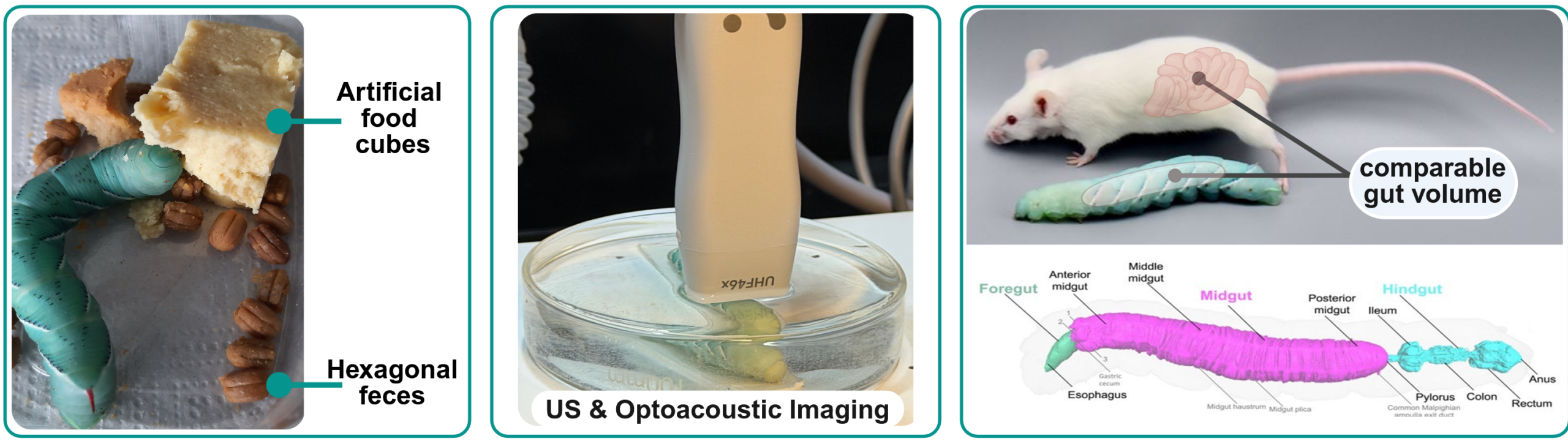
In close cooperation with Dr Anton Windfelder’s research group (https://www.windfelder-lab.com/en), we utilise tobacco hornworm caterpillars (Manduca sexta) as an innovative alternative animal model. Although these insect larvae are evolutionarily distant from humans, both the structure of their intestinal epithelium and the cellular innate immunity of the gut exhibit a strong evolutionary similarity to those of mammals. This makes them a valuable translational model for investigating the fundamental immunology and pathophysiological mechanisms underlying inflammatory bowel disease at the cellular level.
The established intestinal inflammation model of these insect larvae enables the investigation of pathophysiological processes in an ethical and efficient manner.¹,² Building on this, we employ this model organism to test, optimise, and validate modern imaging techniques—with a particular focus on optoacoustics—as well as dyes and contrast agents. As a member of the Erlangen 3R Initiative (https://www.fpz.med.fau.de/manduca-sexta-tabakschwaermer), we make an active research contribution to the animal-welfare-oriented establishment of alternative methods to classical laboratory animals.
1 WINDFELDER, Anton G., et al. High-throughput screening of caterpillars as a platform to study host–microbe interactions and enteric immunity. Nature Communications, 2022.
2 WINDFELDER, Anton G., et al. An enteric ultrastructural surface atlas of the model insect Manduca sexta. iScience, 2024.

Link to study: https://doi.org/10.1016/j.isci.2024.1094102/2